
Importance of OPG X-Ray in Dental Treatment
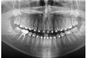
An Orthopantomogram (OPG) is an essential panoramic 2D X-ray that captures the upper and lower jaws, teeth and surrounding bone structures in a single image. Unlike an intraoral X-ray, which focuses on a small area, an OPG provides a broader view of the mouth and jaws.
Different dental specialists use an OPG for different reasons:
- Orthodontist – Planning Tooth Alignment
An OPG helps the orthodontist:
- Evaluate tooth alignment and position.
- Identify unerupted, missing or extra teeth.
- Detect impacted teeth.
- Assess tooth development before treatment.
- Oral Surgeon – Wisdom Teeth and Jaw Problems
- Assesses the position and angle of wisdom teeth before extraction.
- Shows their relationship with important structures, including the nerve canal.
- Helps identify cysts, tumors and jaw fractures.
- Prosthodontist – Crowns, Bridges and Implants
- Assesses the position and condition of neighbouring teeth before crowns and bridges.
- Evaluates available jawbone and surrounding structures for dental implant planning.
- Helps determine whether further 3D imaging is needed for accurate implant placement.
- Periodontist – Assessing Bone Support
- Evaluates bone loss around the teeth.
- Assesses the supporting bone and structures around tooth roots.
- Helps understand the overall impact of gum disease on tooth support.
- Endodontist – Root Canal Treatment
- Helps identify larger infections and lesions around tooth roots.
- Can detect abscesses or cysts near the root tips.
- Detailed intraoral X-rays may be needed to examine individual teeth more closely.
Main Advantages
- Broad view: Shows the jaws and teeth in one quick scan.
- Relatively low radiation: Provides comprehensive information with relatively low radiation exposure.
- Comfortable: Useful for patients who have difficulty opening their mouth or have a strong gag reflex.
Limitations
An OPG provides an excellent overall view, but it does not offer the fine detail needed to examine individual root canals, tiny fractures or subtle early changes around root tips.
Overlapping bones and anatomical structures can also obscure precise details. When necessary, your dentist may recommend intraoral X-rays or 3D imaging such as CBCT.